When an ear sticks out from the head, the structure most often responsible is not the lobe, the helix, or the canal. It is the antihelix: a curved ridge of cartilage that, when it fails to fold during fetal development, lets the outer rim flare away from the skull. The anti helix is a fold of conchal cartilage running inside the outer rim of the ear, and whether it formed cleanly in the womb largely determines how close the ear sits to the head. That same ridge has lent its name to a piercing, and here the trouble starts: two retailers use “anti-helix piercing” for two different locations. This guide separates the anatomy from the piercing, and the piercing-name confusion from the placement a reader actually wants.
The Lobecraft Editorial Desk compiles anatomy terminology from published morphology standards and cross-references piercing jewelry and healing claims against manufacturer and professional-studio sources. What follows draws only on those.
Quick answer: The anti helix is the Y-shaped cartilage ridge inside the outer rim of the ear, formed when conchal cartilage folds between 12 and 16 weeks of gestation. An underdeveloped fold is the usual cause of prominent ears. As a piercing term, “anti-helix” is ambiguous: one retailer defines it as the snug, through the vertical inner cartilage across from the tragus; another treats it as the forward helix, just above the tragus. Both are slow-healing cartilage piercings in the 6-to-12-month range. Because the name does not fix the location, confirm the exact placement with your piercer before the needle goes in.
Where the Antihelix Sits, and What the Fold Separates

The antihelix is a folding of the conchal cartilage. According to the Elements of Morphology project at the National Human Genome Research Institute, it is a Y-shaped curved cartilaginous ridge arising from the antitragus that separates three features of the outer ear: the concha, the triangular fossa, and the scapha. About two thirds of the way along its course it branches into a superior (posterior) crus and a more sharply folded inferior (anterior) crus.
Those branches matter because they bound the spaces around them. The scapha is the groove between the helix and the antihelix. The triangular fossa is the concavity enclosed by the superior and inferior crura of the antihelix and the ascending part of the helix. If you press a finger into the hollow just inside the top of your ear, you are feeling the triangular fossa, walled on two sides by the crura.
Encyclopaedia Britannica frames it from the opposite direction: the antihelix is an inner, concentric ridge that surrounds the concha, separated from the helix by the furrow called the scapha. The two descriptions agree. One starts from the ridge and names what it divides; the other starts from the central bowl and names the ridge around it.
TeachMeAnatomy adds the functional picture. The auricle is a mostly cartilaginous structure that captures and directs sound waves toward the external acoustic meatus, and within it the antihelix runs parallel inside the helix before dividing into its two crura, described there as the inferoanterior and superoposterior crus. The ridges are not decorative. They channel sound inward.
What the Antihelix Does for Hearing, and Why Protruding Ears Do Not Affect It
The external ear is elastic cartilage under tight skin, with ridges that funnel sound into a passageway and on to the eardrum. Per thePlasticsFella’s external ear anatomy overview, that passage, the external auditory canal, is a 25 mm S-shaped channel that directs sound waves to the tympanic membrane. The helix, concha, and tragus shape the sound that enters it; the antihelix is part of that collecting surface.
This leads to a correction worth making plainly. A prominent or protruding ear is a structural and cosmetic matter, not a hearing one. The Children’s Hospital of Philadelphia states on its protruding ears page that protruding ears do not cause functional problems such as hearing loss. The canal and the eardrum do the hearing; whether the outer ridge lies flat against the skull or flares outward does not change what reaches them. The general anatomy references rarely connect the ridge to the protrusion, so this is the useful takeaway: the antihelix’s shape governs how the ear looks and sits, while sound is handled downstream of it.
How the Antihelical Fold Decides Whether an Ear Lies Flat

The fold is not a static feature; it forms on a schedule. ThePlasticsFella’s prominent ear deformity reference puts antihelical folding between 12 and 16 weeks of gestation, and notes that incomplete folding can produce prominent ear deformity. If that ridge does not form fully, the outer rim it would otherwise hold back is left free to flare.
CHOP describes the same mechanism from the clinical side: in most people, protruding ears are caused by an underdeveloped antihelical fold, and when it does not form correctly, the helix sticks out. ThePlasticsFella frames the cause slightly differently, stating that prominent ears usually lack a defined antihelical fold or have a hypertrophic concha. The two sources are materially aligned on the fold; thePlasticsFella adds the conchal-overgrowth alternative as a second contributor. Read together, they point to one structural idea: the ear sits flat when the inner ridge folds, and flares when it does not.
How common is that? ThePlasticsFella reports that prominent ears affect approximately 5% of the population, making them the most common congenital auricular deformity. That figure is from a single source and is best read as an indicative prevalence rather than a settled epidemiological constant.
Surgeons give the structure a working label. ThePlasticsFella describes an “Antihelix Complex,” an aesthetic subunit made up of the antihelix, its crura, and the triangular fossa, used when planning reconstructive work. Grouping the ridge with the hollow it frames reflects how the fold and its surrounding concavity move together when an ear is reshaped.
Prominent-Ear Thresholds: When an Ear Is Measured as Protruding
Two numbers define the threshold, and they come from different sources that happen to agree on the second. CHOP states that ears sticking out more than 2 cm from the side of the head are considered prominent or protruding. ThePlasticsFella sets the clinical criteria as an auriculocephalic angle exceeding 30 degrees or an auricular projection greater than 2 cm from the mastoid surface.
So the reader has two usable markers:
- Projection: more than 2 cm of standoff from the head (CHOP) or from the mastoid surface (thePlasticsFella). The sources land on the same 2 cm figure.
- Angle: an auriculocephalic angle above 30 degrees, per thePlasticsFella. CHOP does not state an angle, so this threshold rests on the one source.
Neither number is a reason to act on its own. They describe when an ear meets a clinical definition, not whether anything should be done about it. Protrusion carries no functional cost, so the decision is cosmetic and personal. Anything that looks asymmetric, painful, or newly changed in an ear’s shape belongs with a clinician, not a measuring tape.
Infant Ear Molding vs. Otoplasty: A Decision Set by the Clock
If the goal is to reshape a prominent ear, timing drives the method, and the window for the easy route is narrow. In the first weeks after birth an infant’s ear is soft and flexible, and CHOP notes that a protruding ear found this early may be corrected without surgery through ear molding.
ThePlasticsFella explains why that window exists and when it closes. In neonates up to 72 hours old, auricular cartilage stays pliable because of residual maternal estrogens, and splinting maintained for 1 to 2 months can realign the ear’s shape. The cartilage is literally mouldable while the hormone effect lasts; after it fades, the cartilage sets.
Miss that window and the correction becomes surgical. Here the two institutions frame timing differently rather than contradicting each other. CHOP states that otoplasty can be performed as early as 5 to 6 years of age, when ears are almost fully grown, reshaping cartilage to create an antihelical fold that holds the ear closer to the head. ThePlasticsFella anchors timing to growth, noting that by age 3 to 4 the external ear reaches approximately 85% of its adult size, which informs waiting until ear growth is nearly complete. The overlap is real: both want the ear near its final size before surgery. The specific age offered differs by source.
Laid out as a decision:
- First few days to weeks of life: non-surgical molding is possible because neonatal cartilage is pliable (up to 72 hours for the most pliable state); splinting runs about 1 to 2 months.
- Age 3 to 4 onward: the ear has reached roughly 85% of adult size per thePlasticsFella, the growth benchmark surgeons watch.
- Age 5 to 6: the earliest CHOP cites for otoplasty, with ears almost fully grown.
This is information, not a recommendation. The right path and timing for a specific child is a conversation for a pediatric surgeon or physician.
After Otoplasty: The Dressing and Headband Schedule

Recovery from otoplasty is largely about protecting the newly created fold while it holds. CHOP notes that the surgical dressing will typically stay in place for 1 to 2 weeks. The surgical fold needs that covered, undisturbed period before anything presses on it.
ThePlasticsFella outlines the follow-on care: a soft, non-compressive headband worn continuously for the first week, then at night for 2 to 4 weeks, with trauma to the ears avoided for at least 3 to 4 weeks. The headband keeps the ear from being bent backward during sleep while the cartilage settles into its reshaped position. These are published general protocols; the operating surgeon’s own instructions take precedence over anything written here, and any pain, swelling, or discharge that seems out of order after surgery is a reason to call the surgical team.
The “Anti Helix Piercing” Naming Problem: One Term, Two Placements
Now to the piercing, where the confusion is real and worth money. Two reputable sources attach the name “anti-helix” to two different holes in the ear, so the term alone does not tell a piercer where to aim.
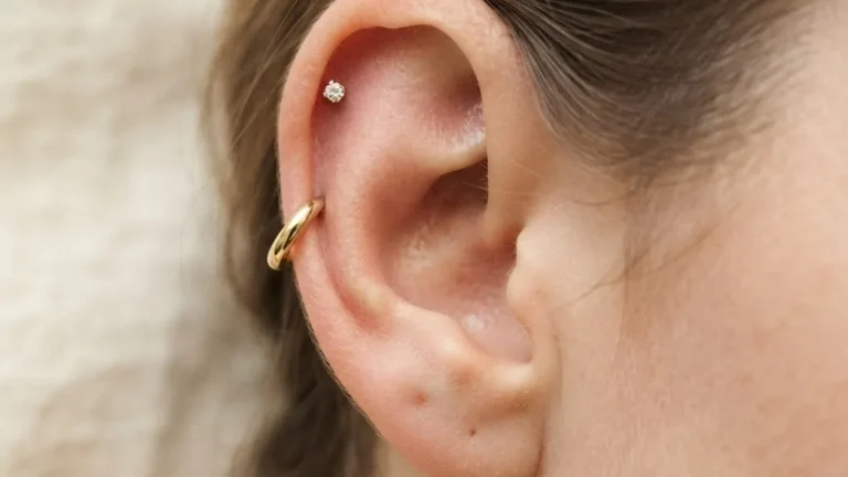
BodyCandy, in its encyclopedia of body piercings, defines the snug (anti-helix) piercing as passing through the vertical inner rim of cartilage generally across from the tragus. That is the snug: a horizontal pass through the antihelical ridge itself, sitting on the inner shelf of the ear.
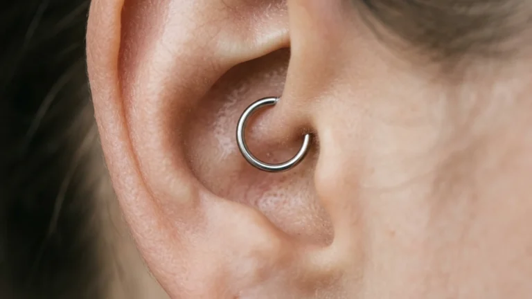
Obsidian Piercing, on its anti-helix piercing page, defines the term differently, treating it as the same as the forward helix, placed near the end of the helix just above the tragus. That is a different structure in a different spot.
These are not two names for one hole. They are two holes under one name, and the sources do not reconcile them. The practical instruction that follows is concrete: do not book an “anti-helix” piercing on the word alone. Point to the exact spot on your ear, or name “snug” versus “forward helix,” and have the piercer confirm the placement before the needle is set. For readers weighing the upper-rim option specifically, our faux rook piercing write-up and the wider set of ear piercings coverage sit nearby.
Anti Helix Piercing Pain and Why the Site Fights You
Whichever placement the name resolves to, the sensation and the slow healing trace back to the same anatomy covered above: this is cartilage, and cartilage is stubborn.
BodyCandy describes the inner-ridge area where the snug sits as one of the most sensitive parts of the ear, and notes that ear cartilage lacks adequate blood supply, making it harder to heal than the lobe. That is the honest pain and healing picture: expect more than a lobe, both in the moment and over the months that follow. Obsidian makes the same vascular point for its placement, stating that ear cartilage is avascular, so antihelix-piercing healing takes longer than the lobe and is more prone to infection.
The reason both sources land on “slower than a lobe” is structural. A lobe is soft, fleshy, and well-supplied with blood that delivers what a healing wound needs. Cartilage is not. With little direct blood flow, the tissue repairs slowly and stays vulnerable to infection for longer. The broader mechanics of this are laid out in our time for cartilage piercing to heal guide.
Healing Windows: 6 to 12 Months, and Why the Ranges Don’t Merge
Both sources quote long healing windows, but they are not describing the same procedure, so they do not combine into a single consensus number.
- BodyCandy (snug, across from the tragus): total healing can be 6 to 12 months or longer.
- Obsidian (its “anti-helix,” placed as a forward helix above the tragus): full healing expected in 6 to 9 months or more.
Because the two describe different placements, these are not two estimates of one healing time. They are healing windows for two different piercings that share a name. Treating them as a single “6 to 9 or 12 month” range would paper over the placement difference that actually determines which one applies to you. Resolve the placement first; the healing window follows from it.
A practical note that holds across both: a cartilage piercing can feel settled on the outside while the channel inside is still open. “It stopped hurting” is not the signal that it has healed. The outer opening closes well before the tract through the cartilage finishes knitting.
Jewelry and Gauge a Piercer Would Start You On
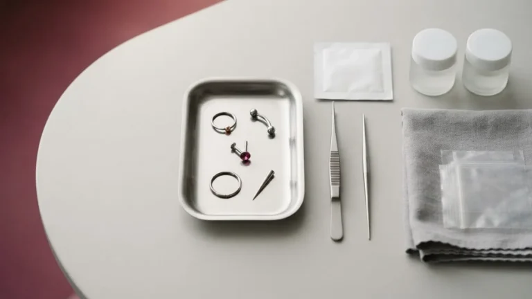
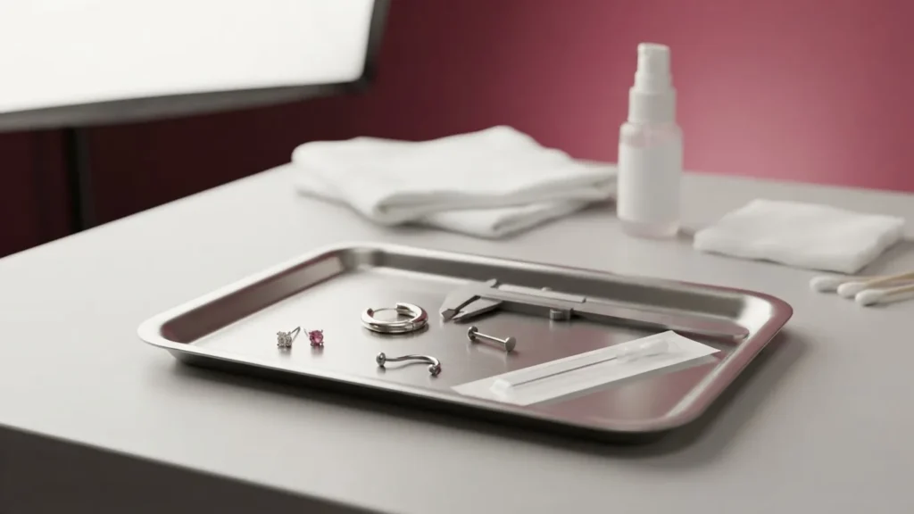
Two points matter for the reader. First, the jewelry style is dictated by the placement. A snug runs horizontally through the inner ridge and takes a barbell that follows that line; a forward-helix-style placement is pierced and fitted differently. This is another reason the naming ambiguity is not academic. Second, the gauge is set by the piercer, not chosen from a catalogue, and heavier or lighter metal is a decision made against your specific cartilage, not a default. Materials and fit should be confirmed against the studio’s own specifications.
When It Is Safe to Change the Anti Helix Jewelry, and the Infection Signs That Say Stop
The timing of a first jewelry change follows the healing window above, not how the piercing looks. Given healing runs 6 to 12 months (snug) or 6 to 9 months or more (the forward-helix placement) by the two sources, an early swap through a channel that is still open inside the cartilage risks tearing the tract and setting healing back. A cartilage piercing that looks closed on the surface can still be fragile within, which is why the months-long window, not the mirror, governs the change. The specifics of judging readiness are covered in the Desk’s changing an ear piercing guide.
Because cartilage is avascular and slower to defend itself, watching for infection matters more here than on a lobe. A piercing that grows more painful, hot, or swollen after the first days, that produces thick or discolored discharge, or that develops spreading redness is signaling a problem, not routine healing. None of that is something to diagnose or treat from an article. The correct move is to see the piercer who did the work, or a clinician, promptly, and to leave the jewelry in unless a professional advises otherwise.
The Anti Helix in One Line: The Fold Behind Both the Ear’s Shape and the Piercing’s Name
Every thread in this guide runs back to the same ridge. The anti helix is the folded conchal cartilage that, when it forms between 12 and 16 weeks of gestation, pulls the outer rim toward the head, and when it does not, leaves the ear prominent in roughly 5% of people. It is part of the surface that funnels sound into the 25 mm canal, which is why its shape is cosmetic rather than a hearing matter. And it is the ridge that gave a piercing its contested name, where “anti-helix” points to either the snug across from the tragus or a forward-helix placement above it, each a slow-healing avascular cartilage piercing, each needing the placement confirmed before anything else. Name the exact spot, respect the months-long healing, and take anything that looks wrong to a professional.
Two Placements, One Name: How Much Longer the Snug Can Take to Heal
The same phrase “anti-helix piercing” points at two different holes, and they do not heal on the same clock. BodyCandy’s snug (anti-helix) is quoted at 6 to 12 months; the retailer that treats “anti-helix” as the forward helix quotes 6 to 9 months. Converting those quoted ranges to weeks (four weeks per month) shows exactly what the naming confusion costs in commitment:
- Snug, worst case: 12 months × 4 = 48 weeks of aftercare before it is fully settled.
- Forward helix, worst case: 9 months × 4 = 36 weeks.
- The penalty for guessing wrong: (12 − 9) × 4 = 12 extra weeks – roughly three additional months of healing if the name turns out to mean the snug rather than the forward helix.
- Shared best case: both start at 6 months × 4 = 24 weeks, so even the quickest outcome is nearly half a year.
- Total uncertainty behind the word: (12 − 6) × 4 = 24 weeks. A single unconfirmed name can hide a six-month swing in how long your ear is a managed wound.
That 12-to-24-week spread is the practical reason to make your piercer name the exact placement before the needle goes in: the label “anti-helix” alone leaves half a year of your healing schedule undecided.
Frequently Asked Questions
Is a snug the same as a forward helix?
Not by anatomy. BodyCandy puts the snug through the vertical inner cartilage across from the tragus; Obsidian Piercing equates “anti-helix” with the forward helix, sitting just above the tragus. Those are separate locations. The label “anti-helix” alone cannot tell you which one a given studio means, so clarify in person.
Can an adult have an otoplasty, or is it only for children?
The cited pediatric guidance gives earliest ages: 5 to 6 years per CHOP, after the ear reaches roughly 85% of adult size by 3 to 4 per thePlasticsFella. Those are lower bounds tied to ear growth, not an upper limit. Candidacy and timing for an older patient are questions for a surgeon, not something these sources settle.
Does an anti-helix piercing affect hearing?
No source links it to hearing loss, and the anatomy argues against it. Sound is processed through the 25 mm canal and the eardrum, downstream of the outer ridges. A piercing through the antihelical cartilage changes the ridge, not the canal. Discomfort or infection is possible; a change in hearing is a separate concern for a clinician.
Why does cartilage healing stretch past a year sometimes?
BodyCandy quotes 6 to 12 months or longer for the snug precisely because cartilage lacks a strong blood supply. Blood carries what a wound needs to close, and the inner ridge gets little of it. The surface can seal while the deeper channel stays open, which is what pushes some healing beyond twelve months.